Skip to content
  • Home
  • About me
  • Clinic
  • Implant-Dentistry
  • Teaching & Learning
  • Courses
  • Resources
  • Home
  • About me
  • Clinic
  • Implant-Dentistry
  • Teaching & Learning
  • Courses
  • Resources
Contact
Implant + Dentistry – Nikos Mattheos
  • Home
  • About me
  • Clinic
  • Implant-Dentistry
  • Teaching & Learning
  • Courses
  • Resources

The roof, the roof, the roof is in fire..! (or shall we say…inflamed!)

  • Nikos Mattheos
  • September 24, 2019
  • 7 Comments
Picture our beautiful 4-storey house. The immaculate building is just delivered to the tenants, still smelling of fresh paint. Nobody notices however a tiny little detail: there is no access to the roof. Soon birds start to gather undisturbed and nest. As time goes by, bird pooh, dust and mud clutters the roof drainage. The recent tropical storm cracked a few tiles and water starts to pool. Soon mosquitos breed by the thousands, flies attract more birds and the roof bursts with life! Not everyone is happy though: first who notices the unbearable stench is the tenant of the Penthouse. But without access to the roof there is nothing to do. A major drama has just unfolded where there is only two possible endings: change the roof, or see all your high-paying tenants moving out one after the other…

Sounds like a very stupid mistake, or? To build a house without access to the roof. Yet this is probably the most common mistake encountered in implant prostheses and the most frequent cause of long-term complications.

by Nikos Mattheos, DDS, MASc, PhD

It’s not the Bacteria, it’s the Biofilm!

There is an inevitable course of events every time a prosthesis is inserted in the mouth. Bacteria, which have always lived in the mouth will contaminate the prosthesis within a couple of minutes. Once some of these bacteria attach themselves on the prosthesis and start to multiply, the prosthesis will be infected. Infection implies the active metabolism and multiplication of bacteria on a surface or tissue. There is nothing wrong with infection, we are infected head to toe and without infection of the oral cavity and the gastro-intestinal tube, we would be simply unable to live. The problem starts however when we allow the infection to mature into a thick, well-structured pathogenic biofilm. This is when infection provokes a response: inflammation. This is the actual disease, in our case “peri-implant mucositis”, introducing a vicious cycle which would sooner or later threaten the very survival of the implant. Biofilm requires two important preconditions: a hard surface to adhere to and a fluid environment with bacteria. The oral cavity is ideal. How certain is it that Biofilm accumulation will lead to peri-implant Mucositis? In my mind, it is as certain as it is that the sun rises from the East.
By now we can cite at least 4 experimental peri-implant mucositis studies in humans (1,2,3,4), all of which demonstrated an undisputable fact: it only takes 2-3 weeks of undisturbed plaque accumulation and every single person developed peri-implant mucositis, no exceptions, none immune. But this does not mean that implants are a lost cause. As we discussed in previous articles, implants are often surrounded by a deep sulcus, which especially in the aesthetic zone can reach 4,5 or even 6 mm. Is this a recipe for trouble? Not necessarily! First, we need to make a clear distinction between a 6 mm periodontal pocket and a 6mm peri-implant sulcus. The first is the result of years of pathology, where the biofilm migrates slowly apically chasing the junctional epithelium. A 6 mm periodontal pocket has an apical migration of the junctional epithelium and pathogenic biofilm established all the way to the bottom of the pocket. The peri-implant sulcus, however is an anatomic structure we can create in an instant as we place the abutment/prosthesis on top of an implant. The prosthesis is free of biofilm and if we can keep it free of biofilm, all evidence points to a long term sustainable health.

A deep sulcus in the interproximal area of anterior implants is the result of the need for natural lookiong aesthetics and paillae. 

can we possibly keep a 6 mm sulcus free of biofilm?
…my response is a clear, Obama-style, “Yes, we can”,
but only if we design it properly.
Some people might now ask, can we possibly keep a 6 mm sulcus free of biofilm? To which my response is a clear, Obama-style, “Yes, we can”, but only if we design it properly. To understand this better, we need to review the mechanism of biofilm formation, which does not start from the bottom of the sulcus, but from the exposed to the mouth hard surface of the prosthesis. In that sense the critical parameter is not if the peri-implant sulcus is 1 or 4 mm deep, but whether we can prevent biofilm accumulation in the very first millimetre, where the prosthesis meets the peri-implant tissues. The fact that Oral Hygiene works is a very good proof of this principle. During the recent experimental mucositis study in Hong Kong (4), when proper oral hygiene was

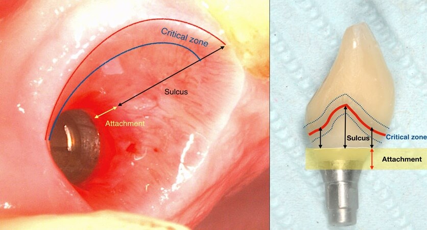
applied at baseline, peri implant tissues with both deep and shallow sulcus remained equally free of inflammation. It is well established since the time of Badersten and Egelberg in the 80’s and 90’s that patient applied oral hygiene will reach no deeper than 0.5-1mm in the sulcus. Yet this was always enough to prevent disease or maintain the outcomes of treatment. So this is where our true battlefield lies: not at the bottom, but at the top of the sulcus, where the prosthesis meets the peri-implant tissues. Disturb the biofilm formation right there, and there will be nothing left to cause inflammation or move apically. Let it accumulate and mature and within a couple of weeks mucositis will be established with the biofilm expanding in all directions, including deeper in the sulcus.

Left: The emergence profile is proper, but the oral hygiene is obviously deficient, with visible deposits of plaque on the crown and an inflamed sulcus.
Right: A proper emergence profile, combined with daily oral hygiene from the patient can maintain the peri-implant tissues free of inflammation, regardless of the depth the sulcus.

It’s not just the Biofilm, it’s the Emergence Profile!

And this is where we reach the “bottom line” of our investigation: What is the single most likely reason for preventing the biofilm from being removed? You guess right: It’s the prosthesis! And actually, not the entire prosthesis, but this critical circumferential couple of mm that we call “emergence profile”. A study in Sweden by Serino et al, found that the prosthesis was blocking the access to oral hygiene in 48% of the patients diagnosed with peri-implantitis. In my experience with such cases, modification of the prosthesis is an essential step before any attempt to decontaminate the implant. What really strikes me though, is that none of the major published studies I have come across so far in the treatment of peri-implantitis actually mentions anything about prosthesis modification in their protocol. Yet it is essential: Treatment of peri-implantitis begins at the prosthesis.

Treatment of peri-implantitis begins at the prosthesis!

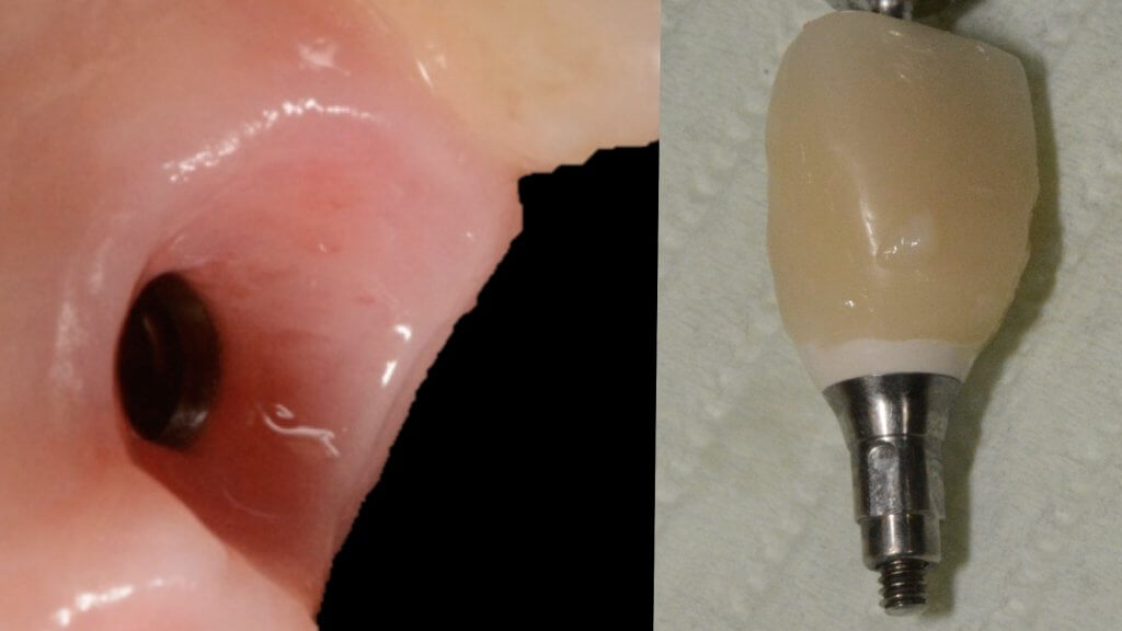
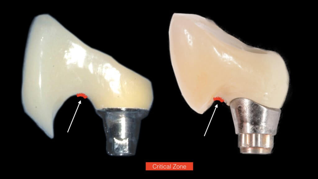
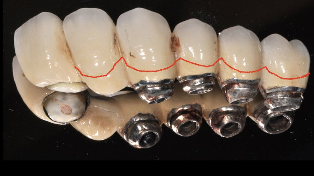
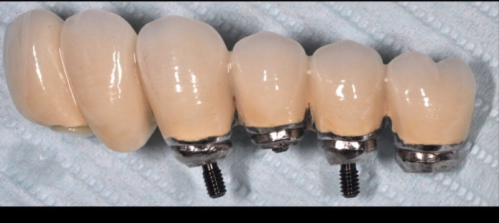
A proper emergence profile is the one that allows direct access to the top of the sulcus and the “danger zone” all around the prosthesis. On the contrary, a typical prosthesis design that blocks access to the sulcus is the “ridge lap”. A ridge lap is a horizontal extension of the prosthesis starting from the top of the sulcus and “riding” the alveolar ridge in mesiodistal or palatolingual direction. We commonly assess the emergence profile in the mesio-distal direction (probably because this is what radiographs show us), but ridge lap is very common (and thus damaging) in the bucco-palatal dimension, too. A recent study by Katafuchi et al, actually showed how the concavity of the emergence profile is important (with concave profile better to convex) and with an emergence angle of  more than 30 degrees being significantly correlated with peri-implantitis. 

But if nobody likes a ridge lap, why do we keep seeing it so often?

There is only one reason: ridge lap is a compensation for compromised  implant selection or position. What are the most common reasons for a ridge lap today:

  • a narrow implant platform selected for the replacement of a wide tooth. This is to be perceived both in mesio-distal and palato-lingual dimensions.
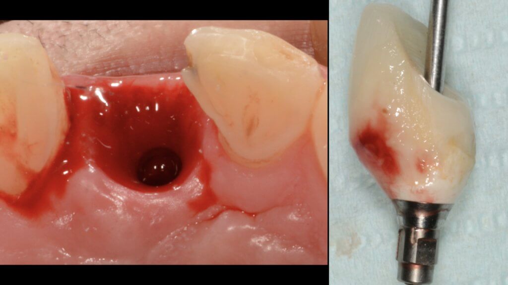
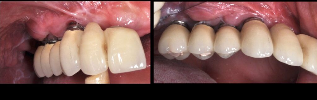
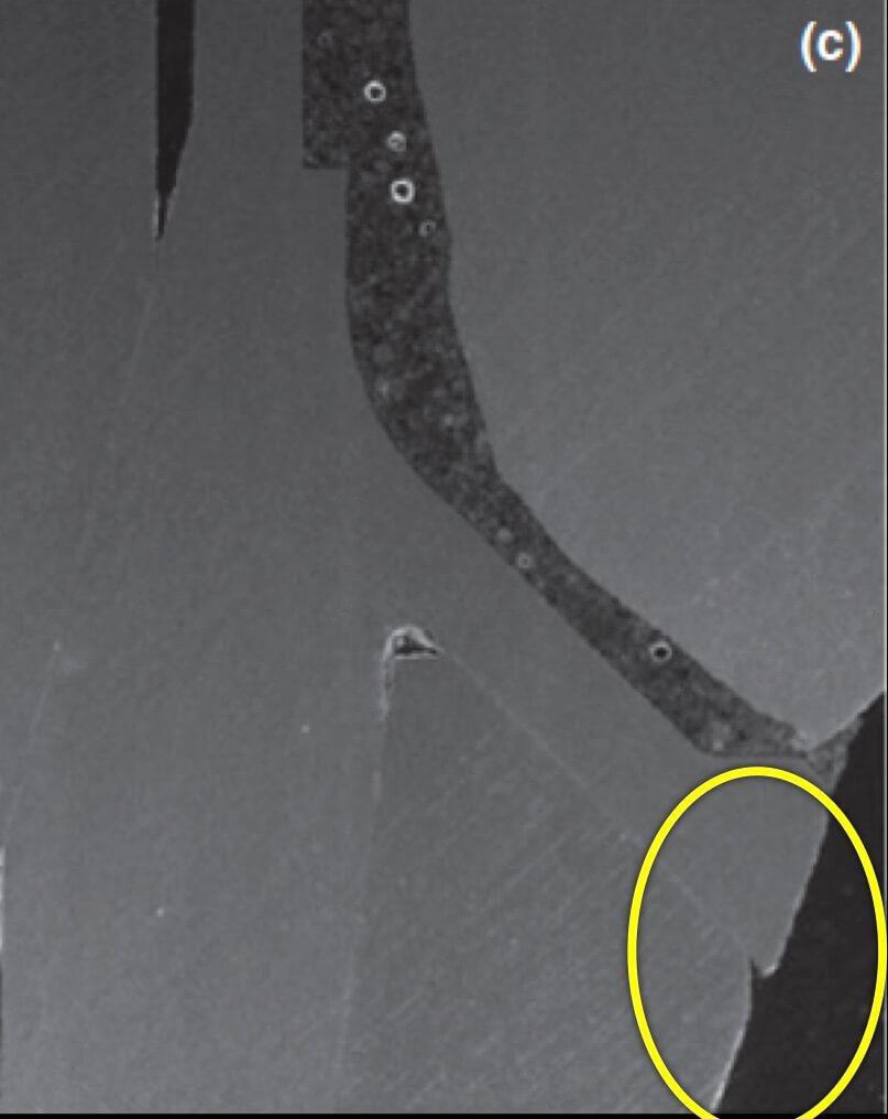
  • too shallow implant placement. This is very commonly encountered in the upper anterior sites, where the implant is typically to be placed below the mesial and distal bone margins (a).
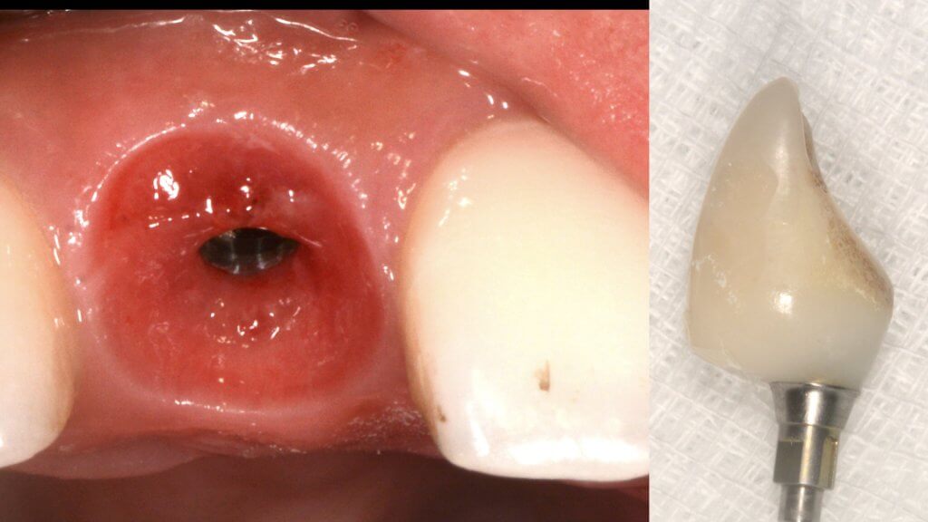
  • too palatal placement, again very common in the upper anterior sites, where the bone resorption pattern eliminates much of the buccal ridge (b) (Photo courtesy Dr. M Janda).
  • too palatal angle of placement (c).
  • wrong mesio-distal position (c).
a. Buccal ridge lap due to shallow placement of anterior implant.
b. Buccal ridge lap due to a combination of too palatal placement and palataly inclined osteotomy
c. Wrong mesio-distal position of the implant predisposing to peri-implantitis

And what if inflammation has already started?

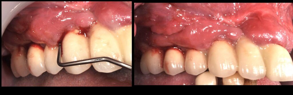
So what if Mucositis kicks in? Well, first thing to do is check the emergence profile: if it blocks access to the sulcus, see if you can modify or change the prosthesis:

Severe Peri-implantitis with swelling and suppuration around uncleanable prosthesis
Complete block of the interdental space due to improper emergence profile
Results after prosthesis modification and 3 weeks of efficient Oral Hygiene. Treatment of Peri-implantitis can now start with surgical disinfection of the implants.
Prosthesis temporarily modified by opening the interdental spaces , trimming excessive convexity and polishing
For more about the management of the above case, click here

Now what if Mucositis is established at sites with deep sulcus?

The experimental mucositis by Chan et al (4) showed that the inflammation burden is greater in the case of a deep sulcus, which could make pathology progress faster or attempts to treat more difficult. However, there was one more particular parameter in this experiment other than the depth of sulcus: the implants used in that study were all tissue level. As opposed to let’s say UCLA abutments, tissue level implants have a significant “gap” and a “notch” exactly in the position where the implant collar meets the crown, which in the case of deep sulcus will be positioned right at the bottom of the sulcus. Does this matter? Well, let’s talk about this more next time …!

Learn the latest news first, subscribe to my newsletter!

References

  1. Pontoriero, et al. Experimentally induced peri-implant mucositis. A clinical study in humans. Clin Oral Implants Res. 1994 Dec;5(4): 254-9.
  2. Zitzmann et al. Experimental peri-implant mucositis in man. J Clin Periodontol. 2001 Jun;28(6):517-23.
  3. Salvi et al. Reversibility of experimental peri-implant mucositis compared with experimental gingivitis in humans. Clin Oral Implants Res. 2012 Feb;23(2):182-90.
  4. Chan D et al. The depth of the implant mucosal tunnel modifies the development and resolution of experimental peri-implant mucositis: A case-control study. J Clin Periodontol. 2019 Feb;46(2):248-255. 7.

Share:

Facebook
Twitter
Pinterest
LinkedIn

7 thoughts on “The roof, the roof, the roof is in fire..! (or shall we say…inflamed!)”

  1. lucy
    September 30, 2019 at 7:58 pm

    its an amazing article can u email it to me
    thank u

    Reply
    • Nikos
      October 11, 2019 at 4:01 pm

      Dear Lucy, thank you for your kind comments! This time I was a bit tight with time and did not make a pdf version of the article, but of course please feel free to download it and save it as a pdf yourself if you want! Again, feel free to share and use for any non-commercial purpose!
      Nikos

      Reply
  2. Matteo
    October 3, 2019 at 11:12 pm

    Very nice article and I like your style. I completely agree with you buy but I think a very important aspect of the inflammation of the tissue around implants is the internal micro leakage effect on the increasing the quantity of anaerobic bacteria in the deepest part of the sulcus (specially in bone level implants). In all the implants/prosthesis connections, existed a gap and a pump like effect of bacteria (from the saliva). The anaerobic are selected to survive in the internal cavity of the system, colonize the tunnel and the Teflon or the cotton or any material the prostodontist use to seal the tunnel.
    That’s why I invented the SilverPlug (as you know). The silver reduce the amount of bacteria inside the system and helps the control of the quality of the biofilm. http://Www.silverplug.ch

    Reply
    • Nikos
      October 11, 2019 at 3:59 pm

      Thank you for your insight! I do understand the problem, which can be a significant threat at times, depending on the implant system and the reconstruction type. In the past for example with older type external connections, this leakage might have been one of the main reasons why bone was always “running away” from the implant shoulder. Having sliced some of the modern internal connections under the SEM, the gap of a bone level with the abutment is less than 2-3 μm which would minimise the possibility of leakage. Then again recently the trend is to use ceramic restorations cemented on titanium “base” abutments, which introduced the risk of a new “gap” in the sulcus, this time between the ceramic crown and the Titanium base… So I think you are right, we need to seriously evaluate the risks of micro-leakage and act accordingly! thank you for your comments!

      Reply
  3. Charles
    October 6, 2019 at 4:43 am

    I love your article Nikos. It paints a very clear picture.

    Reply
    • Nikos
      October 11, 2019 at 3:52 pm

      Many thanks Charles!

      Reply
  4. blog1alex.xyz
    March 19, 2020 at 10:56 pm

    Hi! This post couldn’t be written any better! Reading this post reminds me of my old room
    mate! He always kept talking about this. I will forward this write-up to him.
    Pretty sure he will have a good read. Thank you for sharing!

    Reply

Leave a Comment Cancel reply

Related Posts

  • Featured, Implant Denstistry

Precision Implant Dentistry?

Precision Medicine or P4 is a paradigm of care which considers a patient's individual characteristics to guide diagnosis and treatment. With the wide implementation of a digital workflow, implant dentistry is not fit for P4 practice focused on personalised care, preventive design, predictive algorithms and participatory engagement of the patient.

Read more

  • Nikos Mattheos
  • February 24, 2025
  • 4 Comments
  • Featured, Implant Denstistry

peri-implant mucosa dimensions and emergence profile: a guide for meticulous architects

dental implants placed in fresh extraction sockets were first documented more than 25 years ago, so one might wonder why “immediacy” took so long time to mature into the mainstream implantology. The progress of a scientific protocol however, is rarely a straight line to implementation. Perceived breakthroughs are often followed by backlash and enthusiasm can be at times interchangeable with periods of caution. In the end, technology is also a major catalyst to help a vision make it safe to the mainstream practice. How did immediacy develop from the first “immediate” implants into the current modern concept as a workflow? Let’s take a closer look into a long and winding road!

Read more

  • Nikos Mattheos
  • August 30, 2024
  • 5 Comments
  • Featured, Implant Denstistry

“Immediacy” revisited: beyond the extraction socket!

dental implants placed in fresh extraction sockets were first documented more than 25 years ago, so one might wonder why “immediacy” took so long time to mature into the mainstream implantology. The progress of a scientific protocol however, is rarely a straight line to implementation. Perceived breakthroughs are often followed by backlash and enthusiasm can be at times interchangeable with periods of caution. In the end, technology is also a major catalyst to help a vision make it safe to the mainstream practice. How did immediacy develop from the first “immediate” implants into the current modern concept as a workflow? Let’s take a closer look into a long and winding road!

Read more

  • Nikos Mattheos
  • February 14, 2024
  • 8 Comments
Facebook-f Twitter Youtube Linkedin

About me

  • About Me
  • Teaching & Learning
  • Implant Dentistry

get started

  • Courses
  • Resources
  • Research
  • Feedback

Newsletter

Don’t miss our news and  updates!

  • Home
  • About me
  • Clinic
  • Implant-Dentistry
  • Teaching & Learning
  • Courses
  • Resources